Interactive Diagnosis
You see what we see.
Interactive Diagnosis is the process of showing patients oral conditions during dental exams. Interactive Diagnosis is a fundamental element of dental patient advocacy, supporting the value that we will demonstrate dental conditions before recommending treatment. This is a term Dr. Collins created when he introduced computers to the exam room in 1992.
Key elements of Interactive Diagnosis
Key elements of interactive diagnosis are listening to patient concerns, reviewing medical and dental questionnaires together, sharing digital x-rays on a large computer screen, using video scopes with large computer screens, as well as special macro lens with digital cameras and oddly enough, common hand mirrors. Electronic patient records capture these diagnostic details and make it easier to share them with our patients.
Digital X-Rays
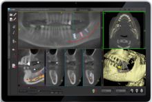
Dentists often used magnifying glasses and special lighting to study traditional x-rays. Now we use digital systems to display x-rays on video monitors during your exam. Features of interest include existing dental restorations, decay or gum disease, and anatomy of the teeth and jaws. Patients are often interested to see how deep an area of decay is, and exactly where it is so effort to brush and floss can be focused where it is needed most. More info about digital x-rays.
Video Scopes show the "Big Picture"
I remember going to a dentist as a child, hoping he wouldn't find anything as he picked through my teeth talking jargon to his assistant. He said I had cavities now and then, but I never saw one, and most of what the dentist did was somewhat mysterious to me. Part of my personal mission is to offer a pleasant, enlightened dental experience in a personal way. Video scopes can be used to demonstrate nearly any dental condition easily. We can capture video images to our electronic patient records.
We have a video scope that is used inside the mouth with an infection control baggie to explore teeth, gums, and other oral features. This is useful for displaying broken teeth, focal areas of gum disease, corroded or worn edges of fillings, and many other conditions. I wish my childhood dentist could have shown me the funny bumps on the back of my upper canines that I sometimes worried about - the bumps were normal anatomy, and I was sure something was wrong!
Digital Photography in Dentistry
We love our Fuji S2 Pro digital camera. Our system was provided by Washington Scientific Camera, with a dedicated macro lens (for close-ups), a matching macro speed flash, and circuits for through the lens flash metering. The photos from this system are high enough quality for publication. We use the camera for before and after photos, pictures of pathologic lesions, and for optimal shade matching with porcelain artists at our laboratory.
Electronic Records in Dentistry
We use computers to build electronic patient records, complete with tooth charts, pictures from our video scope, digital x-rays, and more. We print treatment plans that include diagnostic details related to each procedure in the plan. Dr. Collins was a pioneer in computing in dentistry, adopting his first dental software system in 1983. He was a charter member of the Task Force for Dental Informatics, created by the American Dental Association to create standards for electronic records in dentistry in the early 1990's. He went on to create systems for teledentistry and digital attachments for electronic insurance claims. You can find more details in Dr. Collins Curriculum Vitae page.
 by Keith Collins, DMD
