

3D Cone Beam Imaging
A quick, comfortable scan that shows what flat X-rays can't
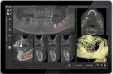
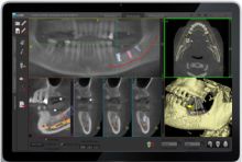
A standard dental X-ray is a flat picture. It can hide the width of the bone, the path of a nerve, or a problem sitting behind a tooth. Cone beam computed tomography (CBCT) captures your teeth, jaws, nerves, and sinuses in three dimensions. We can then view any area from every angle and in thin cross-sections, and build custom panoramic and 3D views for each case.
Cone beam imaging is a dental version of the medical CT scan. A CT scan uses a flat, "fan-shaped" beam. Cone beam uses a "cone-shaped" beam, like a teepee, that captures the whole area in a single rotation. The result is fine detail with a much shorter scan time and a fraction of the radiation of a medical CT scan. Cone beam X-rays are easy for the patient and surprisingly affordable.
What to Expect
Open, quick, and painless
- No tunnel. The scanner is open. You stay still while the arm makes one pass around your head.
- Fast. The scan itself takes less than a minute.
- Nothing to prepare. We simply ask you to remove earrings, glasses, and removable dental appliances.
- See it together. Dr. Collins reviews your 3D images with you on screen, part of our Interactive Diagnosis approach, so you understand your options.
How 3D Imaging Helps
Better information means more predictable treatment
- Dental implant planning - measure bone height and width, and locate nerves and sinuses before surgery.
- Impacted and wisdom teeth - see exactly where a tooth sits relative to nerves and neighboring roots.
- Root canal problems - find extra canals, cracks, and infections that flat X-rays can miss.
- Sinus and bone evaluation - assess the sinus floor and bone before grafting or implant placement.
- Unexplained pain or swelling - look for cysts, lesions, and other hidden causes.
Dual Scan Imaging
Combining images of the patient and the planned teeth
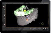
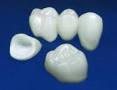
Dual scan imaging merges a standard cone beam image of the jaws with a second 3D scan of the planned teeth, such as crowns, a bridge, or a denture. This enables optimal planning for the most complex cases.
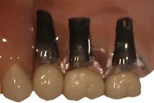
The purpose of implant planning is to create a surgical plan that supports the final teeth. In other words, we plan where the teeth will be before we plan where the implant "roots" will go.
3D Surgical Guides for Dental Implants
Guides are "printed" from our cone beam implant plan
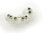
Our 3D planning software connects directly with the dental lab. Once we finish the design for any number of implants, the lab produces a surgical guide. It snaps onto the remaining teeth or fits a jaw that has no teeth.
The lab "prints" the guide in a medical-grade polymer. The guide transfers the plan precisely to the mouth, and detailed planning cuts the time needed for implant placement by about half.
Questions About 3D Imaging?
Dr. Collins has been placing dental implants since 1986
If you're considering implants or have a problem that ordinary X-rays haven't explained, a cone beam scan may give the answer. Call us at (360) 574-7477 or request an appointment.